CFA FCT/UNL is one of the two scientific groups of Centro de Física Atómica (CFA). Most of its members belong to Faculdade de Ciências e Tecnologia da Universidade Nova de Lisboa (FCTUNL).
Study of lead accumulation in bones of Wistar rats by X-ray fluorescence analysis: aging effect
| Title | Study of lead accumulation in bones of Wistar rats by X-ray fluorescence analysis: aging effect |
| Publication Type | Journal Article |
| Year of Publication | 2012 |
| Authors | Guimarães, D., Carvalho M. L., Geraldes V., Rocha I., and Santos J. P. |
| Journal | Metallomics |
| Volume | 4 |
| Pagination | 66-71 |
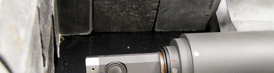
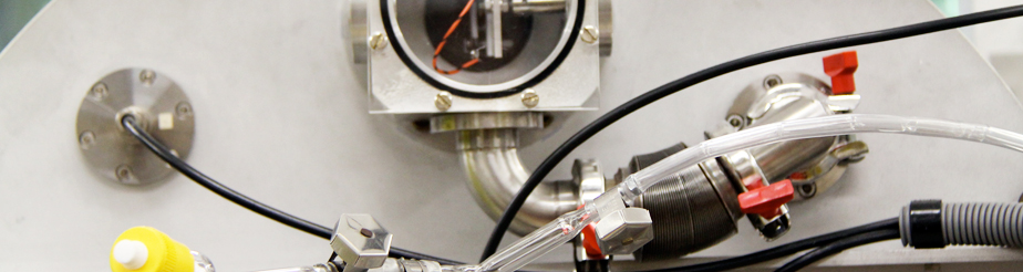
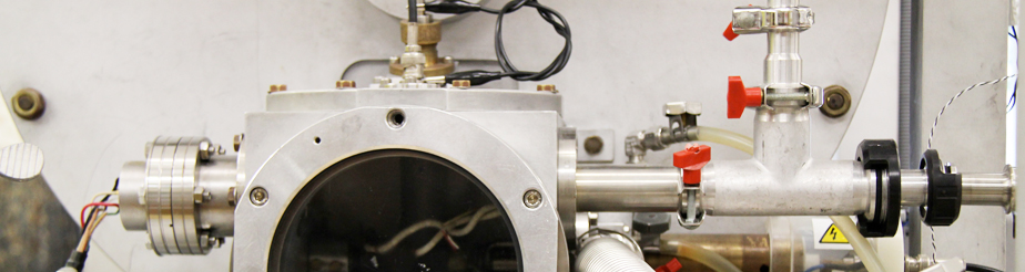
| Abstract | The accumulation of lead in several bones of Wistar rats with time was determined and compared Q3 for the different types of bones. Two groups were studied: a control group (n = 20), not exposed to lead and a contaminated group (n = 30), exposed to lead from birth, first indirectly through mother’s milk, and then directly through a diet containing lead acetate in drinking water (0.2%). Rats age ranged from 1 to 11 months, with approximately 1 month intervals and each of the collections had 3 contaminated rats and 2 control rats. Iliac, femur, tibia–fibula and skull have been analysed by energy dispersive X-ray fluorescence technique (EDXRF). Samples of formaldehyde used to preserve the bone tissues were also analysed by Electrothermal Atomic Absorption (ETAAS), showing that there was no significant loss of lead from the tissue to the preservative. The bones mean lead concentration of exposed rats range from 100 to 300 mg g 1 while control rats never exceeded 10 mg g 1. Mean bone lead concentrations were compared and the concentrations were higher in iliac, femur and tibia–fibula and after that skull. However, of all the concentrations in the different collections, only those in the skull were statistically Q4 significantly different (p o 0.05) from the other types of bones. Analysis of a radar chart also allowed us to say that these differences tend to diminish with age. The Spearman correlation test applied to mean lead concentrations showed strong and very strong positive correlations between all different types of bones. This test also showed that mean lead concentrations in bones are negatively correlated with the age of the animals. This correlation is strong in iliac and femur and very strong in tibia–fibula and skull. It was also shown that the decrease of lead accumulation with age is made by three plateaus of accumulation, |
| URL | http://dx.doi.org/10.1039/c1mt00149c |
Research